-
 Brian Kelly
★★★★★
in the last week
Brian Kelly
★★★★★
in the last week
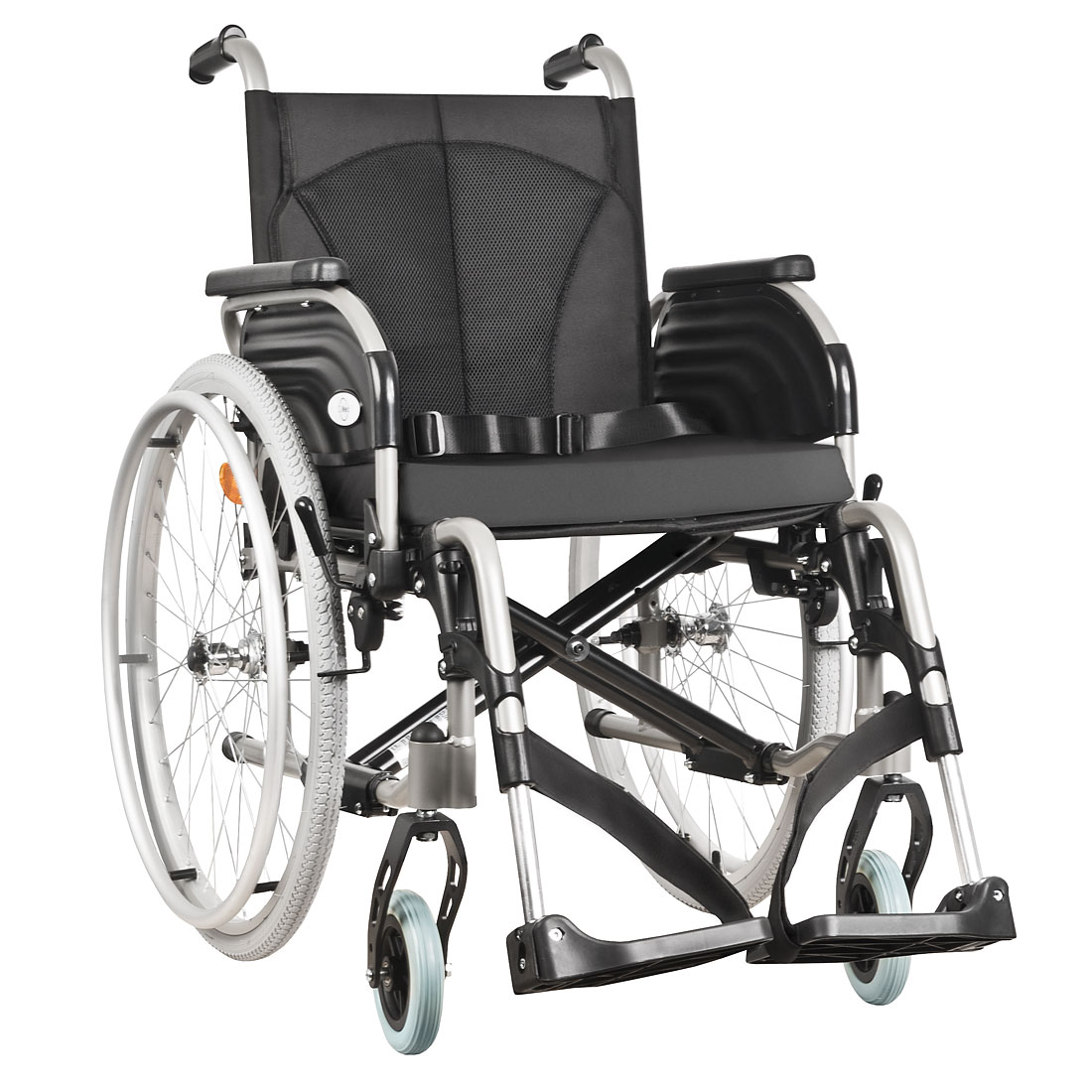
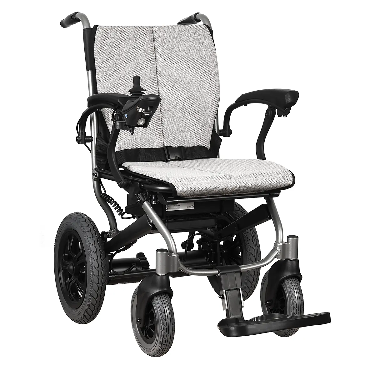
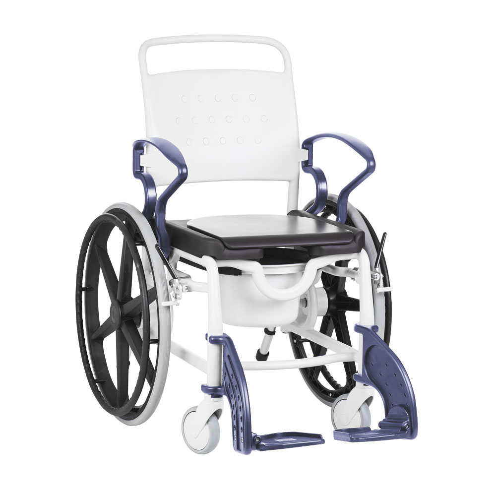
Thanks heaps for the quick delivery and awesome little wheel chair.The colour also made it a bit more happier for my mum , she just loved it. Great quality and price.
-
 Glenn and Annette Hyland
★★★★★
2 weeks ago
Glenn and Annette Hyland
★★★★★
2 weeks ago
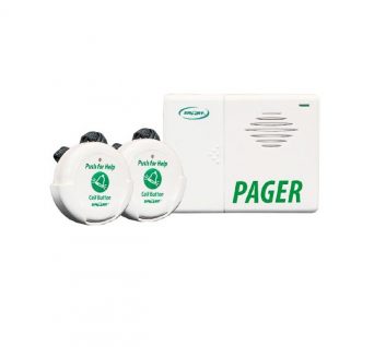
The call button we purchased has been great. As a person with a disability, I can use it daily to call my carer wherever she is in the house.Only issue is that the sound is very loud and cannot be adjusted.Suits … More our needs.
-
 Deb GT
★★★★★
2 weeks ago
Deb GT
★★★★★
2 weeks ago
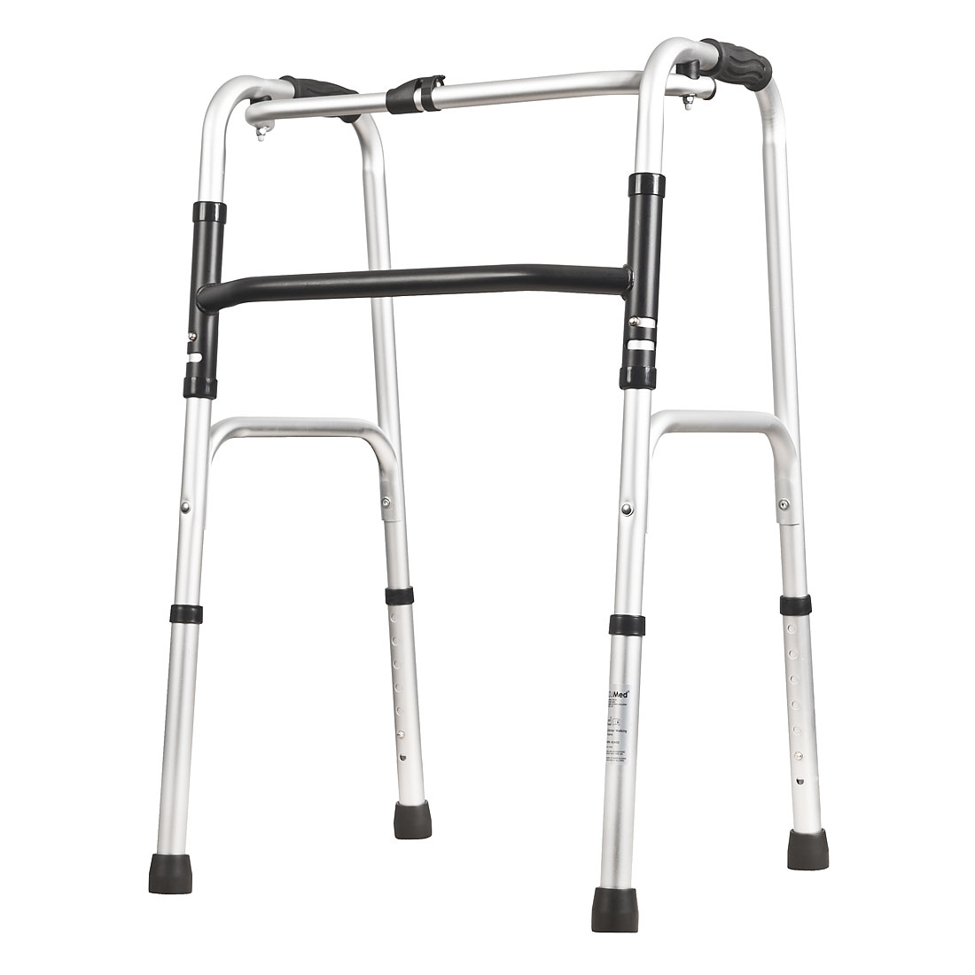
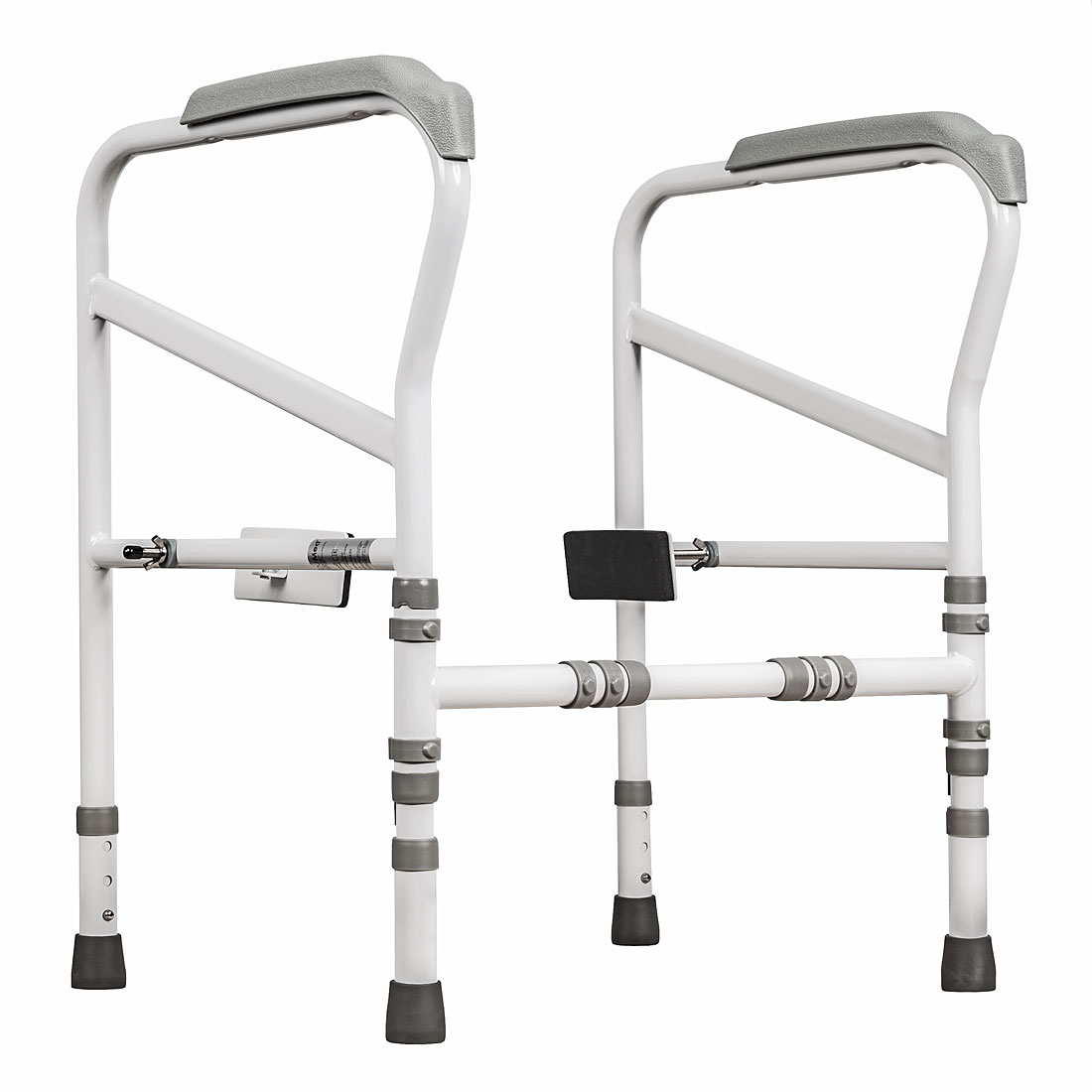
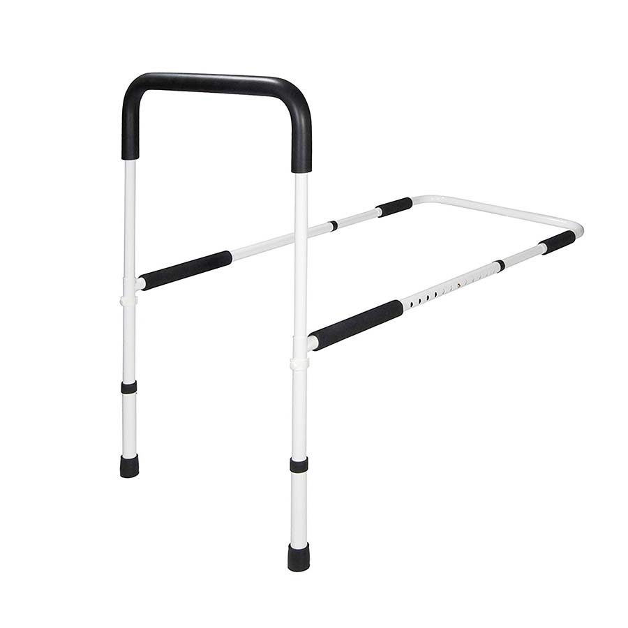
My 97 year old Mum really appreciated the bed rail. She usually complains about anything I buy her. This time she has only positive feedback which is great. Thanks heaps. Cheers Deb
-
 Karen Clark
★★★★★
2 weeks ago
Karen Clark
★★★★★
2 weeks ago
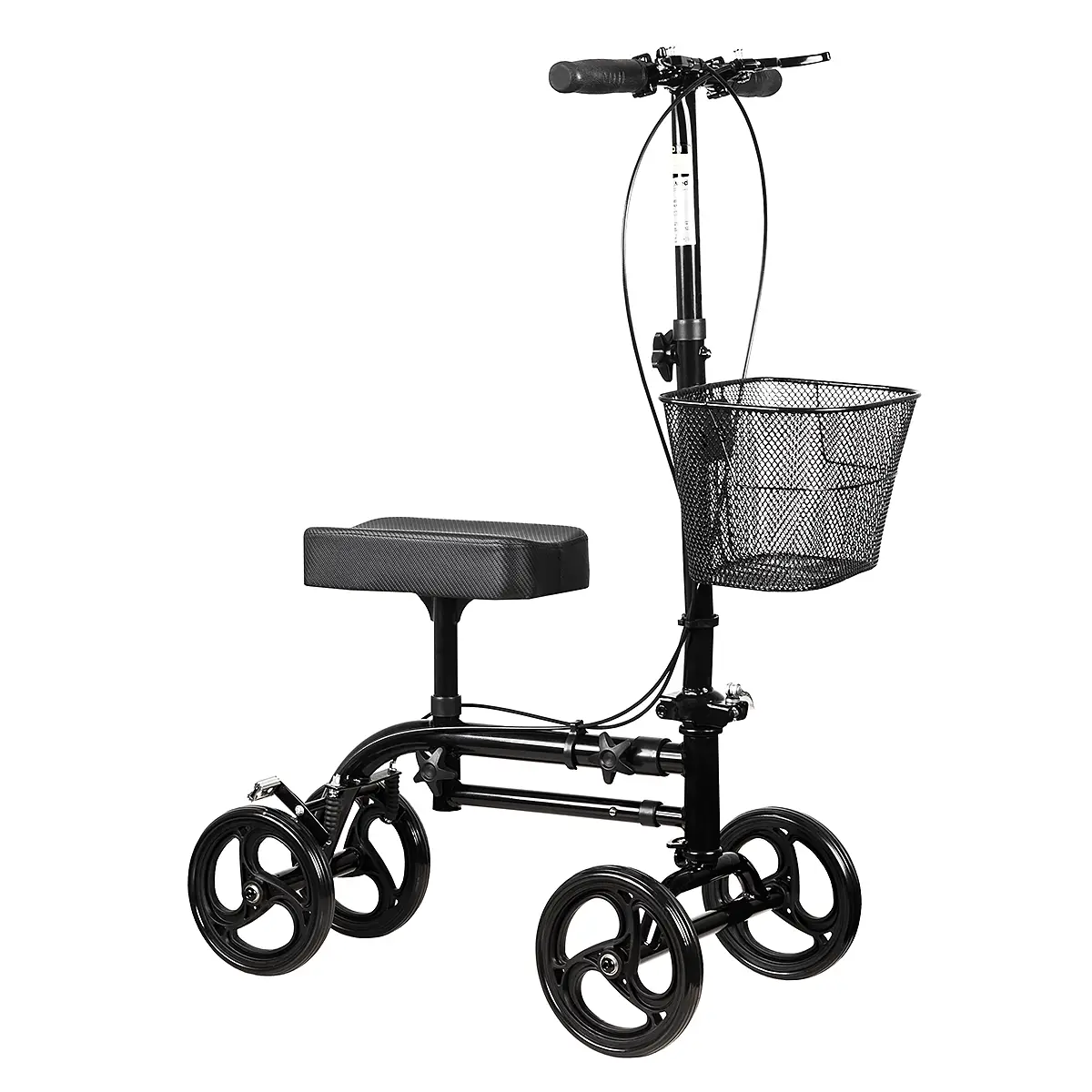
Ordered the Knee Scooter & it arrived promptly. The scooter worked well both indoors & outside. Very comfortable knee pad. The attached bag was also very useful.
-
 Diana Nick
★★★★★
2 weeks ago
Diana Nick
★★★★★
2 weeks ago
This was my second order—very happy with the quality and fast shipping. Will definitely order again. Highly recommended!